The Challenge
- Membrane-bound targets sometimes cannot be made into recombinant protein for screening
- Some recombinant forms of membrane-bound targets do not present accurate structure
The Single Cell Solution
- Develop screening on cell surface of target-expressing cells
- Combine cell-based screens (SurfScreen) with recombinant screens (SimulScreen), when possible, to enrich data
Highlights
- Predicted cell-binders and non-binders confirmed by flow cytometry
- Target-binding to cells between 13 pM to 1 nM
- Sequence lineages show the same phenotype in the screen
Problem: Taking on the Challenge
AbTheneum efficiently screens antibody binding profiles with recombinant proteins, yet some membrane targets require screening with cells due to their structure.
SurfScreen is a new module for AbTheneum for screening antibodies against membrane targets.
Solution: Finding a Better Way
Wild-type mice were immunized with TIGIT protein. Antibody-secreting cells were isolated using CD138 positive selection. The CD138+ cells were deposited onto a picowell device and aligned with a second device loaded with both TIGIT-expressing cells and Parental cells expressing GFP. The two devices were pressed
together aligning all picowells, allowing antibodies from CD138+ cells to interact with TIGIT and Parental cells (Figure 1).
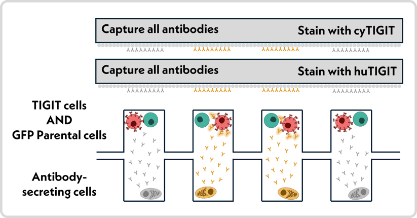
Figure 1. Schematic of SurfScreen project, screening all secreted antibodies against TIGIT-expressing cells, Parental cells, recombinant human TIGIT protein, and recombinant cynomolgus TIGIT protein.
Want the movie trailer, not the whole film?


The two devices were separated, the TIGIT cells were stained with fluorescent secondary antibody to detect cell-binding antibodies. Two antibody arrays were captured from the CD138+ cells and stained with recombinant human and cynomolgus TIGIT protein (Figure 1, gray rectangles) All CD138 cells were lysed antibodies captured for sequencing using AbTheneum sequence capture protocols. Clonotypic antibodies show similar protein and cell binding profiles as expected (Figure 2).
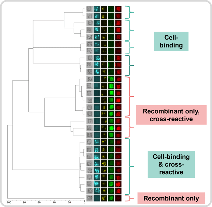
Figure 2. Antibody sequences from a SurfScreen campaign on TIGIT, showing sequence similarity on a dendrogram and images from the screen: Brightfield, Cell-binding signal (cyan), Parental cell signal (yellow), Recombinant hTIGIT (green), Recombinant cyTIGIT (red). Related antibody clonotypes show similar phenotypes.
Twelve (12) diverse mAbs were selected from the output—2 non-cell binders and 10 cell-binders—and validated by Flow against TIGIT and Parental cells (Figure 3). All 12 mAbs were negative against Parental cells (not shown), and all 10 cell-binders showed high affinity against TIGIT cells, EC50 between 13 pM to 1 nM. The 2 non-cell binding antibodies showed no binding to TIGIT cells, confirming the predictive power of the assay.
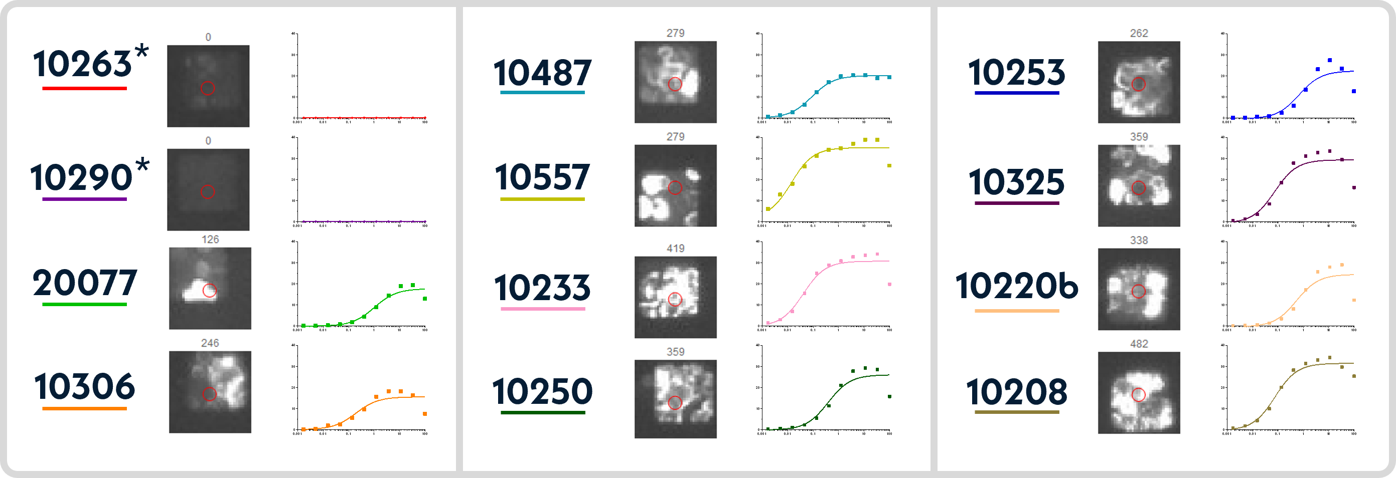
Figure 2. Twelve antibodies expressed, 2 non-cell binding* and 10 cell-binding. Each mAb presented with cell-binding signal captured via SurfScreen and Flow cytometry plot against TIGIT-expressing cells.
Impact: What We Achieved
SurfScreen can combine recombinant and cell-based screens for membrane-bound targets with high predictive power in the screening data. Multi-layered screening that includes cell-binding can help discover differentiated antibodies.
SurfScreen is also being used for multipass membrane targets like GPCRs and using virus-like particles in the AbTheneum workflow.
Curious what this could look like for your program?